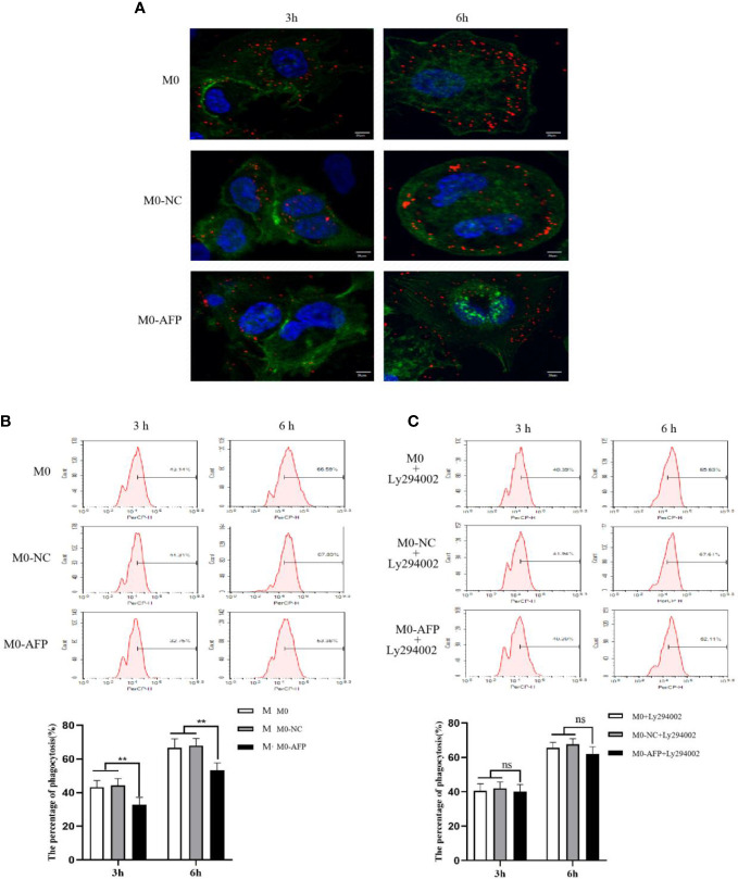
Figure 5.
The effects of AFP and Ly294002 (PI3K inhibitor) on macrophages enggulfing polystyrene latex beads. M0 macrophages were infected with an AFP-expressing negative control lentivirus vector(M0-NC) or AFP-expressing lentivirus vector(M0-AFP) for 48 h and then treated with LPS+IFN-γ for 24 h to induce M0 macrophage polarization into M1-like phenotype. (A) Macrophages engulfed polystyrene latex beads in the M0, M0-NC and M0-AFP groups at 3 h and 6 h were observed by laser confocal microscopy; the fluorescence intensity represents the numbers of phagocytoses. Blue: cell nucleus(DAPI stained); Green: cytoplasm(5-(and -6)-carboxyfluorescein diacetate succinimidyl ester(CFSE) incorporation of the intracellular fluorescent dye); Red: polystyrene latex beads. (B) Macrophages engulfed the numbers of polystyrene latex beads in the M0, M0-NC and M0 AFP groups at 3 h and 6 h, detected by flow cytometry; the lower column diagram shows the statistical analysis of the phagocytosis rate of each group, **P<0.01. (C) After treatment with the PI3K inhibitor Ly294002 for 24 h, macrophages engulfed the numbers of polystyrene latex beads in the M0, M0-NC and M0-AFP groups at 3 and 6 h were detected by flow cytometry; the low column diagram shows the statistical analysis of the phagocytosis rate of each group, P>0.05. The images are from the at least three independent experiments.

