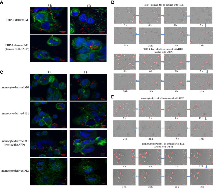
Figure 7.
The effect of tAFP on macrophages phagocytizing polystyrene latex beads or HCC cells. Monocytes from health donors were treated with PAM to induce the polarization into M0 macrophage, then M0 macrophages were administered with LPS+IFN-γ or IL-4+IL-13 for 24 h to induce M0 macrophage polarize towards M1-like phenotype or M2-like phenotype(showed in Supplementary materials: S-Figure 2 ). The THP-1 derived M1-like macrophage(gray) or health monocytes derived macrophages(gray) co-cultured with polystyrene latex beads(red) or HLE cells(red), then treated with tAFP(final concentration 20μg/mL). (A) THP-1 derived M1-like macrophage co-culture with polystyrene latex beads and treated with tAFP, the images of macrophages phagocytize polystyrene latex beads were taken by laser laser confocal microscopy after co-culture for 3h or 6h. Blue: cell nucleus(DAPI stained); Green: cytoplasm(CFSE incorporation of the intracellular fluorescent dye); Red: polystyrene latex beads. (B) THP-1 derived M1-like macrophages co-cultured with HLE cells and treated with tAFP, the macrophages phagocytosis was observed by an intelligent living-cell high-throughput imaging analyzer, the bright field channel was used to photograph, and images were taken every 30 min for 24 h. (C) Health monocytes-derived macrophage co-cultured with polystyrene latex beads and treated with tAFP, the images of macrophages phagocytize polystyrene latex beads were taken by laser laser confocal microscopy after co-culture for 3h or 6h. (D) Health monocytes derived M1-like macrophages co-cultured with HLE cells, then treated with tAFP, the M1-like macrophages phagocytosis was observed by an intelligent living-cell high-throughput imaging analyzer, the bright field channel was used to photograph, and images were taken every 30 min for 24 h. The images are from the at least three independent experiments.

