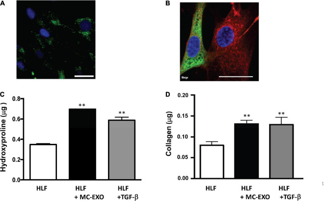
FIGURE 1.
MC-EXO are taken up by HLFs and stimulate collagen synthesis. (A) Representative epifluorescence image of HLF uptake of MC-EXO labeled with PKH-67 (green). Nuclei are labeled with 4′,6-diamidino-2-phenylindole (DAPI) (blue). Scale bar = 15 μm. (B) Representative confocal image of HLF uptake of MC-EXO labeled with CellVue Claret Far Red (red). HLFs were transiently transfected with mNeonGreen-KDEL, a fluorescent marker of the endoplasmic reticulum. Nuclei are labeled with DAPI (blue). Note that only the HLF on the left was successfully transfected with the mNeonGreen-KDEL marker. Scale bar = 15 μm. (C) Hydroxyproline content of lysates from HLF, HLF incubated with MC-EXO (40 μg total protein) or TGF-β (10 ng/ml) at the 72 h time point. All assays performed in triplicate and normalized to total protein (μg). **P < 0.01 versus HLF, n = 5 samples/group. (D) Secreted collagen in supernatants of HLF, HLF incubated with MC-EXO (40 μg total protein) or TGF-β (10 ng/ml) for 72 h. All assays were performed in triplicate and normalized to total protein (μg). **P < 0.01 versus HLF, n = 5 samples/group.

