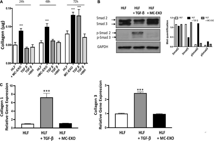
FIGURE 3.
Uptake of MC-EXO by HLF stimulates collagen synthesis via a Smad-independent pathway and with different kinetics from TGF-β. (A) MC-EXO (40 μg total protein, black bars) and TGF-β (10 ng/ml, dark gray bars) stimulated collagen secretion in HLFs after 24, 48, and 72 h and in untreated HLFs. Shown is the inhibition of TGF-β stimulated collagen secretion in HLFs by the TGF-βR1 inhibitor SB525334 (TGF-βR1 INH, 10 μM, light gray bars). All assays performed in triplicate and normalized to total protein (μg). ***P < 0.001 versus HLF, n = 6 samples/group. (B) Western blot of HLF lysates at 24 h and after treatment with TGF-β (10 ng/ml) or MC-EXO (40 μg total protein). Smad 2/3 and p-Smad 2/3 show signaling via the TGF-β pathway. GAPDH is loading control. Western blot was performed twice with associated quantification. (C) Collagen 1 and collagen 3 mRNA expression in HLFs and HLFs treated with either TGF-β or MC-EXO analyzed by RealTime qPCR and normalized to GAPDH mRNA. ***P < 0.001, n = 4 samples/group.

