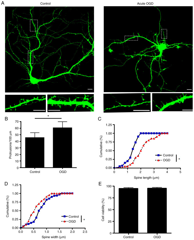
Figure 1.
Acute OGD treatment impairs dendritic spine morphology. (A) Primary hippocampus neurons were transfected with enhanced green fluorescent protein. Upper images represent neurons treated with OGD and visualized under confocal microscopy at DIV17 (scale bar, 10 µm). Lower images are magnified image from the doted square regions of the upper image. (B) Number of protrusions. Spine (C) length and (D) width (n=1,200-1,500 spines from ≥20 neurons). (E) High-density cortical neurons were treated with OGD at DIV17 and tested with MTT (n=3 batches of neurons). *P<0.05. OGD, oxygen-glucose deprivation; DIV, days in vitro.

