
Figure 1.
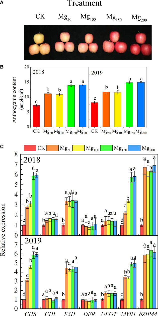
Effects of Mg application on fruit peel coloring. A representative photograph (A) shows the color of the fruit peel under different Mg treatments. Anthocyanin concentration (B) and anthocyanin biosynthesis-related gene expression (C) of fruit peel were measured in each treatment. The error bars indicate SD of six replications. Significant differences were detected by different letters at P ≤ 0.05.