FIGURE 1.
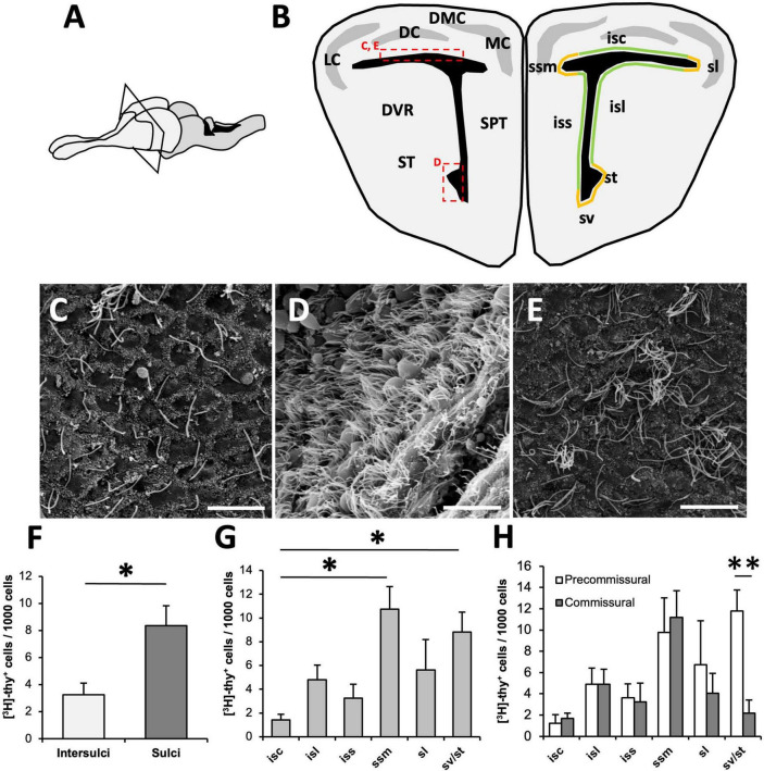
Organization of the ventricular zone (VZ) in Podarcis liolepis. (A) Schematic of the brain of P. liolepis. The telencephalon is represented in a lighter shade. (B) Diagram of a transverse section of the telencephalon at an intermediate pre-commissural level. In the left hemisphere are represented the main telencephalic regions, while in the right hemisphere shows the regionalization of the VZ in sulci (orange) and intersulcal regions (green), divided as sulcus septomedialis, sulcus lateralis, sulcus ventralis, and sulcus terminalis. Insets indicate the approximate location of the images shown in panels (C–E). (C) Scanning electron microscopy (SEM) image showing the surface of the dorsal wall of the lateral ventricle (LV). In this region most cells are uniciliated. (D) SEM image of the surface of the LV at the level of the sv/st where most cells are multiciliated. (E) SEM image of the LV dorsal wall surface, in which some clusters of multiciliated cells intermingled with uniciliated cells can be observed. (F) Regional distribution of [3H]-thymidine-positive ([3H]-thy+) cells 1.5 h after [3H]-thymidine administration. Proliferative cells are more frequently found in sulci than in intersulcal regions (n = 5, two-tailed paired t-test). (G) Graph showing the distribution of [3H]-thy+ cells after 1.5 h of survival within the different sulci and intersulcal regions of the LV. The sulcus septomedialis and the sulci ventralis/terminalis presented a significantly higher number of proliferating cells than the intersulcus corticalis (n = 5, Friedman’s test followed by Dunn’s multiple comparisons test). (H) Distribution of [3H]-thy+ cells after 1.5 h of survival in the different sulci and intersulcal regions comparing pre-commissural and commissural levels. Proliferating cells were equally distributed in anterior and posterior levels in all regions except in the sulci ventralis/terminalis, with a higher concentration of [3H]-thy+ cells in anterior levels than in caudal levels (n = 5, two-tailed paired t-test). DC, dorsal cortex; DMC, dorsomedial cortex; DVR, dorsal ventricular ridge; isc, intersulcus corticalis; isl, intersulcus lateralis; iss, intersulcus septalis; LC, lateral cortex; MC, medial cortex; sl, sulcus lateralis; SPT, septum; ssm, sulcus septomedialis; ST, striatum; st, sulcus terminalis; sv, sulcus ventralis. Data are represented as the mean ± SEM. *p < 0.05, **p < 0.01. Scale bars: 10 μm.

