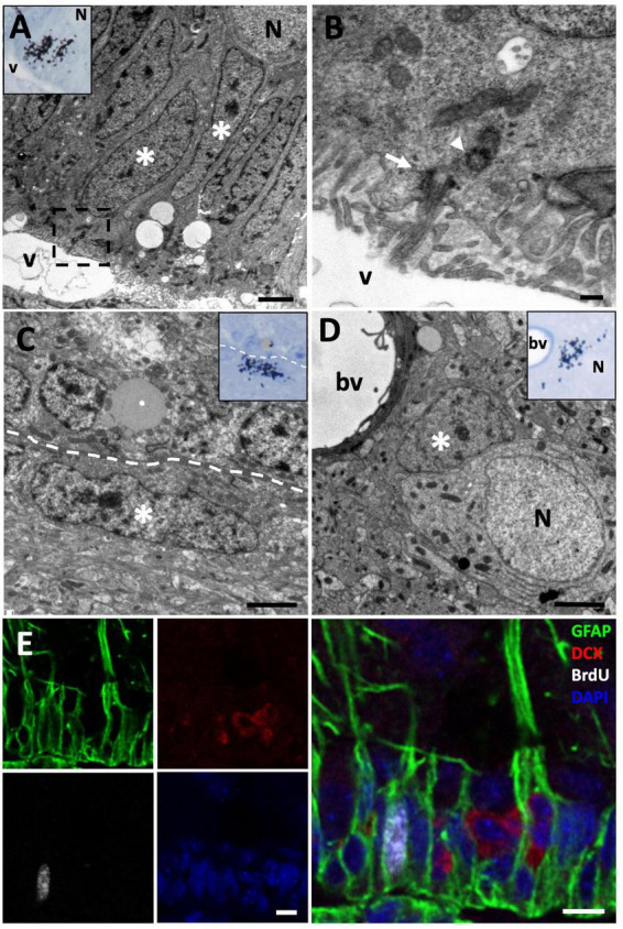
FIGURE 2.

[3H]-thymidine-incorporating type B cells but not neuroblasts are found in the ventricular zone (VZ) at short survival times (1.5 h). (A) [3H]-thymidine-positive ([3H]-thy+) radial glia cells (asterisks) were identified in the sulcus medialis of the (VZ) in animals with survival times of 1.5 h. (B) Detail of the inset in the previous image, showing the contact of this type B cell with the ventricular lumen and the presence of a primary cilium (arrow) and its associated daughter centriole (arrowhead). (C) [3H]-thy+ B cell after 1.5 h survival in an intersulcal region. These cells generally present a flattened morphology. (D) [3H]-thy+ cell with glial features located in the brain parenchyma. (E) Immunofluorescence detection in the anterior olfactory nucleus of an animal injected with bromodeoxyuridine (BrdU) with a survival time of 1.5 h, in which BrdU+/GFAP+ cells are found, whereas all DCX+ neuroblasts at this time-point were BrdU–. bv, blood vessel; N, neuron; v, ventricle. Scale bars: (A,C,D) 2 μm; (B) 200 nm; (E) 5 μm.
