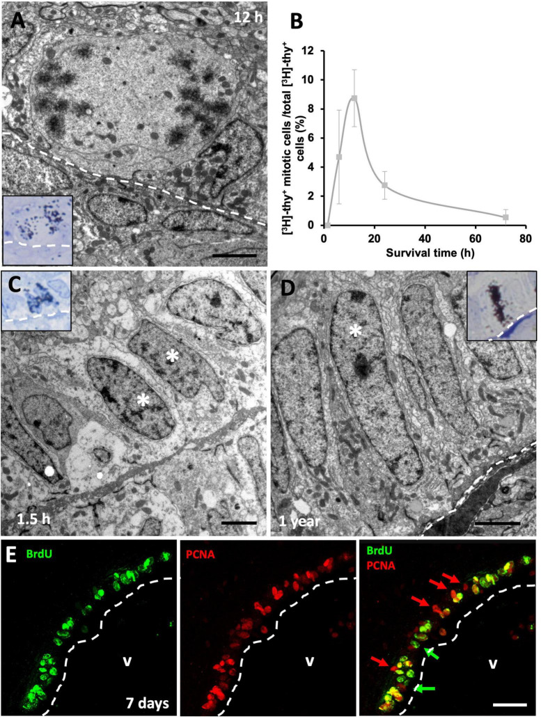
FIGURE 4.
Proliferative rate of neural progenitors in the ventricular zone (VZ). (A) [3H]-thymidine-labeled ([3H]-thy+) mitotic cells in contact with the lateral ventricle (LV) lumen were quantified 12 h following [3H]-thymidine administration. (B) Quantification of the percentage of [3H]-thy+ cells in mitosis relative to all [3H]-thy+ cells in the VZ 1.5, 6, 12, 24, and 72 h after [3H]-thymidine administration. (C) Doublet of non-mitotic [3H]-thy+ cells (asterisks) in the 1.5 h survival group. (D) Slow-dividing [3H]-thy+ type B cell (asterisk), corresponding to a uniciliated radial glia in contact with the LV lumen, 1 year after [3H]-thymidine administration. (E) BrdU/PCNA immunofluorescence detection in the sulcus medialis of an animal injected with BrdU with a survival time of 7 days. Most cells are double-positive, although some BrdU–/PCNA+ and some BrdU+/PCNA– cells were also detected. Scale bars: (A–C) 2 μm; (E) 25 μm.

