FIGURE 6.

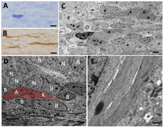
Tangential migration of neuroblasts in the olfactory peduncle. (A) Longitudinal section of the olfactory peduncle stained with toluidine blue. (B) Consecutive section to that shown in panel (A) on which post-embedding immunohistochemistry for DCX was performed. Several DCX+ cells with elongated morphology can be observed. (C) Transmission electron microscopy (TEM) image of the region shown in panel (A). The DCX+ cells shown in panel (B) can be observed (asterisks). (D) Neuroblasts (with their nuclei labeled with an A) migrate over radial glia cells (labeled with B) that line the ventricle of the olfactory peduncle and are also in direct contact with adjacent neurons (N). (E) Detail of several neuroblasts processes sectioned longitudinally with presence of microtubules. Scale bars: (A,B) 5 μm; (C,D) 2 μm; (E) 500 nm.
