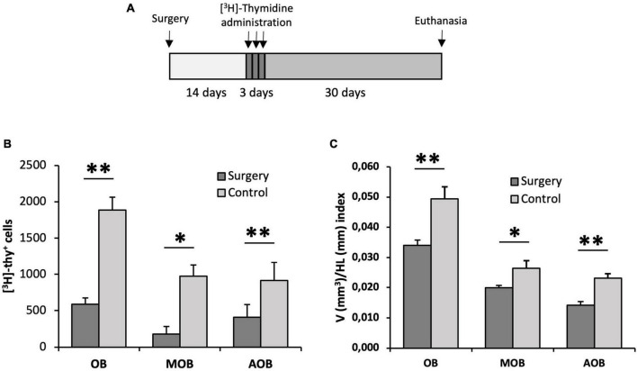
FIGURE 7.
Proliferation assessment in the olfactory bulb of animals with their olfactory peduncle sectioned. (A) Surgery and [3H]-thymidine administration protocol. (B) Quantification of the total number of [3H]-thymidine-positive cells found in the olfactory bulb (OB), main olfactory bulb (MOB) and accessory olfactory bulb (AOB) using the physical dissector. Cell proliferation in all regions was significantly reduced in lizards with their olfactory peduncles sectioned compared with control animals (n = 5, two-tailed unpaired t-test). (C) Cavalieri estimation of the volume of the OB, MOB, and AOB relative to the head length in the surgery and control groups. This volume/head length index was significantly lower in the surgery group (n = 5, two-tailed unpaired t-test). Data are represented as the mean ± SEM. *p < 0.05, **p < 0.01.

