FIGURE 8.

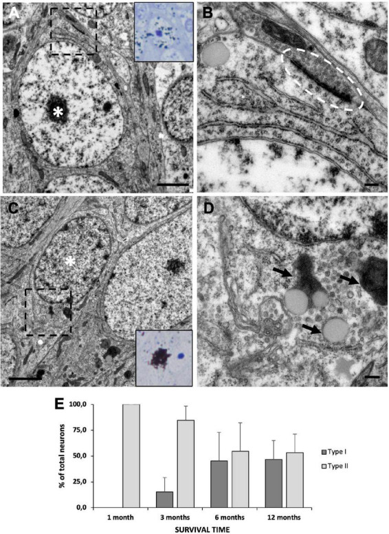
Neuronal maturation in the medial cortex (MC). (A) MC neuron labeled with [3H]-thymidine after 1 month of survival following administration (asterisk). It presents large nucleus with relaxed chromatin and a prominent nucleolus, and electron-dense cytoplasm with abundant free ribosomes, typical features of type II neurons. (B) Detail of a synapsis (dotted oval) established by a type II neuron. (C) MC neuron labeled with [3H]-thymidine after 1 year of survival following administration (asterisk). It presents a small nucleus with clumped chromatin and electron-lucent cytoplasm, typical of type I neurons. (D) Detail of lipofuscin aggregates (arrows) in a type I neuron. (E) Quantification of the percentage of type I and type II [3H]-thymidine-positive neurons at different survival times after administration. In the MC, the percentage of type I neurons increases with time, while that of type II neurons decreases accordingly. Data are represented as the mean ± SEM. Scale bars: (A,C) 2 μm; (B,D) 200 nm.
