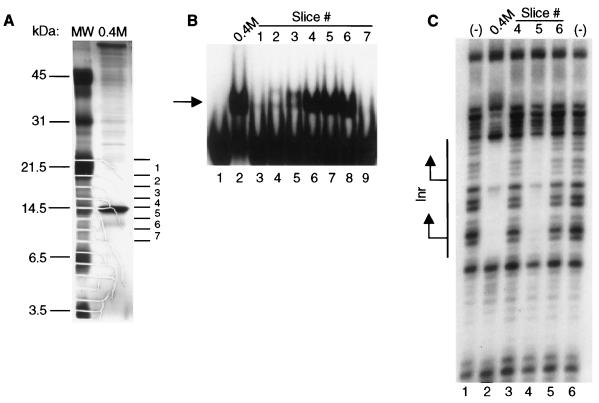
FIG. 3.
The 14.5-kDa protein band contains the Inr-binding activity. (A) Protein from the 0.4 M KCl final Inr column fraction was precipitated with 25% TCA and separated in duplicate lanes on a 17.5% Tris-glycine SDS-PAGE gel. Gel slices, ca. 4 mm in length, were excised from one lane for the denaturation-renaturation analysis, while the remainder of the gel was silver stained. The approximate position of each gel slice is indicated to the right of the silver-stained gel, and the molecular mass markers are shown on the left. (B) Gel shift assay with the −15/+15 αSCS Inr probe (Fig. 1) and either the 0.4 M KCl fraction (lane 2) or an equal volume of the eluate recovered from the individual gel slices following the denaturation-renaturation protocol (lanes 3 to 9). The Inr-specific binding activity is indicated by the arrow. (C) DNase I footprinting assay performed with either no protein (lanes 1, 6), the 0.4 M KCl fraction (lane 2), or an equal volume of the eluate recovered from gel slices 4, 5, and 6 (lanes 3 to 5, respectively) and the wild-type αSCS Inr probe. The location of the Inr element is shown with the two transcription start sites indicated by the arrows.

