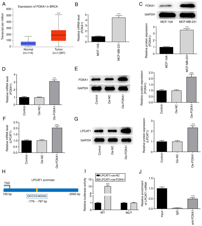
Figure 4.
Association between FOXA1 and LPCAT1. (A) UALCAN database analysis based on The Cancer Genome Atlas data indicates that FOXA1 is highly expressed in breast cancer tissues. ***P<0.001 vs. normal. Expression levels of FOXA1 in MCF-10A and MDA-MB-231 cells were assessed using (B) RT-qPCR and (C) western blotting. ***P<0.001 vs. MCF-10A. Expression of FOXA1 in the MDA-MB-231 cells transfected with oe-FOXA1 was confirmed using (D) RT-qPCR and (E) western blotting. Expression of LPCAT1 in the transfected MDA-MB-231 cells was confirmed using (F) RT-qPCR and (G) western blotting. ***P<0.001 vs. oe-NC. (H) Binding sites for transcription factors FOXA1 and LPCAT1 promoters predicted using the HumanTFDB website. (I) LPCAT1 promoter activity was determined with a luciferase reporter assay. ***P<0.001 vs. LPCAT1 + oe-NC (WT). (J) Binding between FOXA1 and LPCAT1 was evaluated with a chromatin immunoprecipitation assay. ***P<0.001 vs. IgG. FOXA1, forkhead box A1; LPCAT1, lysophosphatidylcholine acyltransferase 1; RT-qPCR, reverse transcription-quantitative PCR; HumanTFDB, Human Transcription factor Database; oe, overexpression; NC, negative control; BRCA, breast invasive carcinoma; WT, wild type; MUT, mutant.

