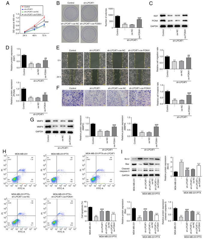
Figure 5.
FOXA1 regulates LPCAT1. (A) Proliferation and (B) colony formation of MDA-MB-231 cells co-transfected with sh-LPCAT1 and oe-FOXA1 was assessed. (C and D) Expression levels of Ki67 and PCNA were determined using western blotting. (C) Representative images and (D) densitometrically quantified results are presented. (E) Cell migration and (F) invasion potential were assessed using wound healing and Transwell assays, respectively. Scale bar, 100 µm. (G) Expression levels of MMP2 and MMP9 were determined using western blotting. ***P<0.001 vs. control; ##P<0.01 and ###P<0.001 vs. sh-LPCAT1 + oe-NC. (H) Apoptosis after 4 nM PTX treatment was assessed by flow cytometry. (I) Enrichment of apoptosis-associated proteins in the co-transfected cells was determined using western blotting. ***P<0.001 vs. MDA-MB-231; ###P<0.001 vs. MDA-MB-231/PTX + sh-NC; ΔΔΔP<0.001 vs. sh-LPCAT1 + oe-NC. FOXA1, forkhead box A1; LPCAT1, lysophosphatidylcholine acyltransferase 1; sh, short hairpin; oe, overexpression; NC, negative control; PCNA, proliferating cell nuclear antigen; OD, optical density; PI, propidium iodide; FITC-A, fluorescein isothiocyanate-Annexin V.

