
Figure 1. Combining imaging with cell sorting enables new experimental strategies across the life science disciplines.
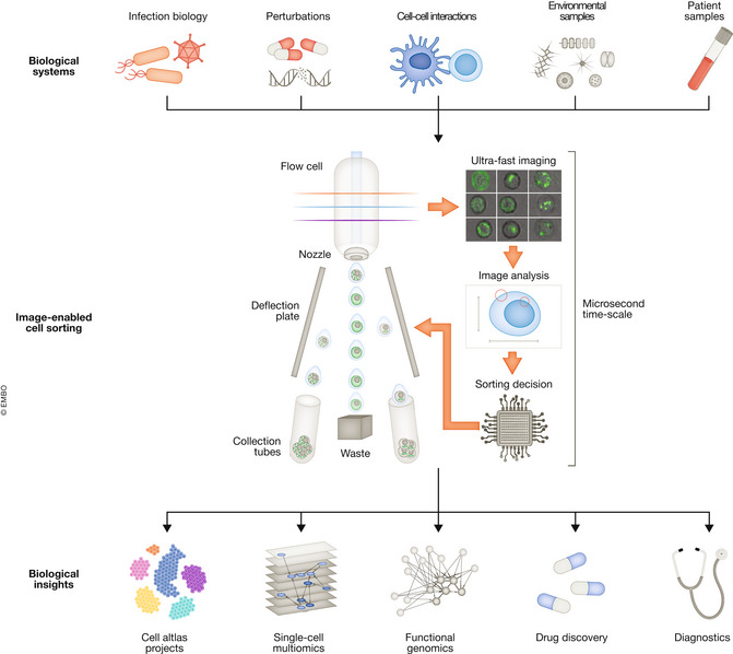
Examples of prime applications for image‐enabled cell sorting. Top: Image‐enabled cell sorting isolates cells from multiple sample sources, such as in vitro model systems that were genetically perturbed, treated with drugs, or infected with bacteria or viruses. In addition, dissociated tissues, patient biopsy material, and environmental samples can be used to isolate spatial phenotypes of interest. Middle: Separation according to spatial microscopic phenotypes by classifying events according to phenotypic measurements or machine learning‐based methods. Bottom: Sorting using image information identifies novel cell types and states for cell atlas projects, is used for single‐cell multi‐omics (see main text), or enables high‐speed functional genomic screening. Cells with specific phenotypic properties, such as production cell lines that generate drugs, or highly active T‐cells that preferentially interact with antigen‐presenting cells, are isolated. Information from images will be used for the fast diagnosis of diseases.