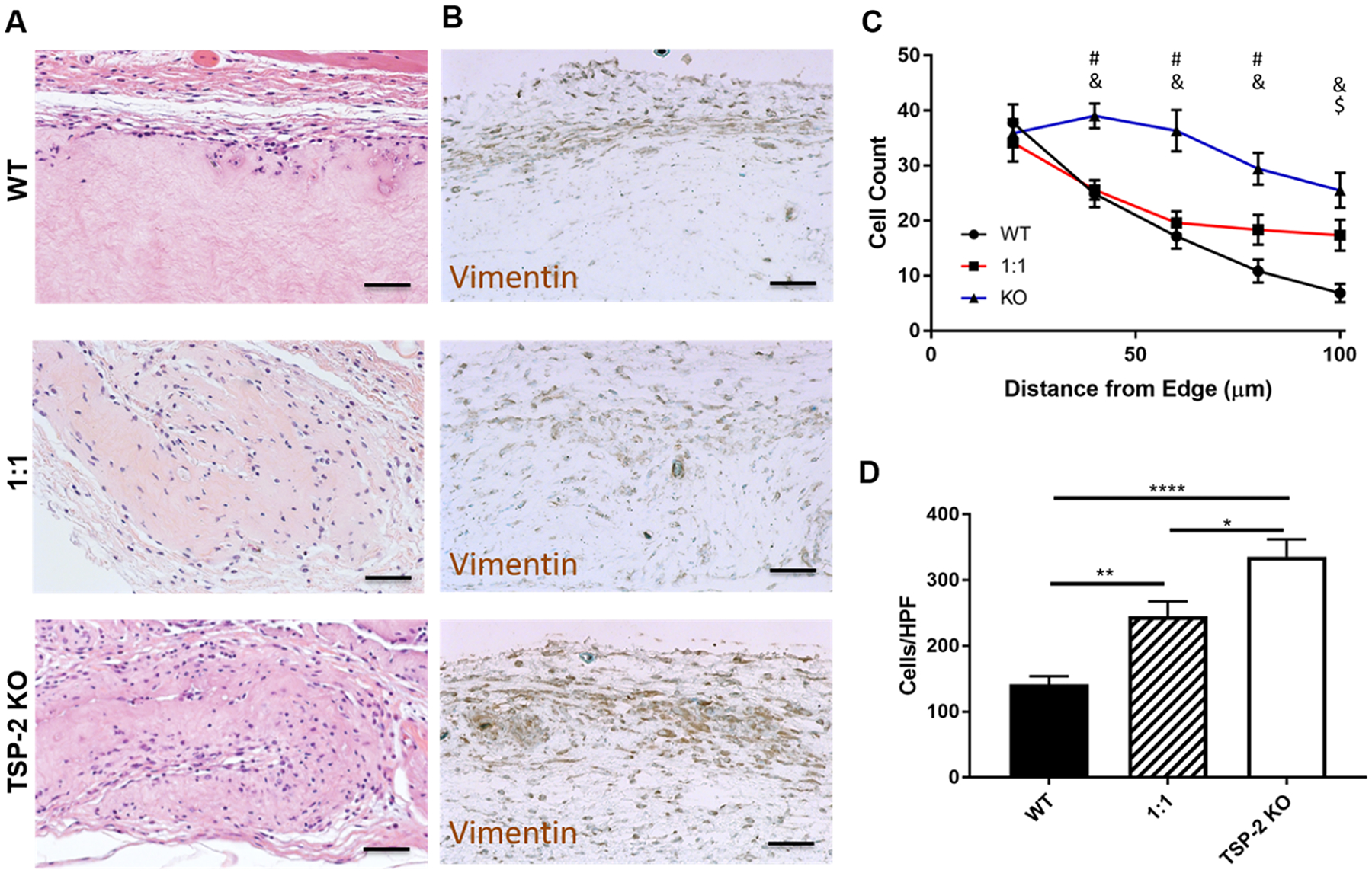
Figure 3.

Genetic manipulation permits tunability of cell invasion into tissue-derived hydrogels. (A) Representative H&E images indicate higher cell presence in TSP-2 KO gels that were implanted subcutaneously in healthy mice for 5 days. (B) Vimentin staining indicates that many of the cells present were of mesenchymal lineage. (C) Quantification shows that cells penetrated further into TSP-2 KO hydrogels and (D) an increasing ratio of TSP-2 KO matrix in the hydrogel resulted in increased total cellular content. Scale bars = 50 μm. Results are given as mean + SEM [±SEM for (C)], n = 8, *p < 0.05, **p < 0.01, ****p < 0.001. # indicates that TSP-2 KO is different from WT, $ indicates that 1:1 is different from WT, and & indicates that 1:1 is different from TSP-2 KO, p < 0.05.
