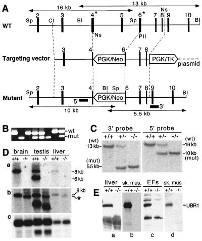
FIG. 2.
Construction of UBR1−/− mice. (A) Top diagram, a restriction map of the ∼24-kb 5′-proximal region of the ∼120-kb mouse UBR1 gene. Middle diagram, the targeting vector. Bottom diagram, the deletion and/or disruption UBR1− allele. Exons are denoted by solid vertical rectangles. The directions of transcription of the neomycin (neo) and the thymidine kinase (tk) genes are indicated. Homologous recombination resulted in the replacement of the UBR1 exons 4 to 6 with the neo cassette. Exon 4, marked by an asterisk, contains Gly147 and Asp150, the residues that are essential, in S. cerevisiae UBR1, for binding type 1 destabilizing N-terminal residues (see Results). Exon 6, also marked by an asterisk, contains Asp233 and His236, the residues that are essential, in S. cerevisiae UBR1, for binding type 2 destabilizing N-terminal residues (see Results). Probes used for Southern hybridization are indicated by solid rectangles. Restriction sites: Sp, SphI; Cl, ClaI; BI, BamHI, Ns, NsiI, PII, PvuII. (B) PCR analysis of mouse tail DNA. The primers were 5′-GCCACTTGTGTAGCGCCAAGTGCCAG-3′ (for neo; forward), 5′-GAGATAGGAAACTGCATGCGCTGC-3′ (for UBR1; forward), and 5′-CAAGAGTGCAACAGTTACCACATG-3′ (for UBR1; reverse). DNA bands corresponding to the wild-type (wt) and mutant (mut) UBR1 alleles are indicated on the right. (C) Southern analysis of BamHI-cut (3′ probe) and SphI-cut (5′ probe) tail DNA from +/+, UBR1+/−, and UBR1−/− mice. The 3′ probe detected 13- and 5.5-kb UBR1 fragments in BamHI-cut DNA corresponding to the wild-type and mutant UBR1 alleles, respectively. The 5′ probe detected 16- and 10-kb fragments in the same SphI-cut alleles. The depicted organization of the deletion and/or disruption UBR1−/− allele was additionally verified using Southern analysis with other restriction endonucleases (data not shown). (D) Northern analysis. Total electrophoretically fractionated RNA from brain, testis, or liver of +/+ and UBR1−/− mice was probed either with the UBR1 cDNA fragment (nucleotides 555 to 888) which was deleted in the UBR1−/− allele (gel a) or with the ∼2-kb cDNA fragment (nucleotides 116 to 2124) that contained both the deleted region (nucleotides 532 to 885) and its flanking sequences (gel b) or with the human β-actin cDNA fragment (gel c). (E) Immunoblot analysis of total extracts from liver, skeletal muscle, and EFs with affinity-purified antibody (38) against the N-terminal ∼35-kDa fragment of mouse UBR1 (gels a to c) or with affinity-purified antibody specific for the 2-1 UBR1 peptide (see Materials and Methods) encoded by the deleted region of UBR1 in the UBR1− allele (gel d).

