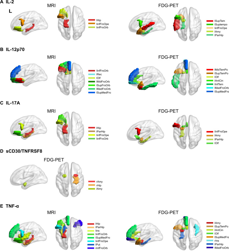
Fig. 2.
Brain regions associated with peripheral inflammation markers. Graph depiction of the brain regions significantly correlated with peripheral inflammation markers, including A IL2; B IL-12p70; C IL-17A; D sCD30/TNFRSF8; and E TNF-α. The brain regions are mainly distributed in frontal–temporal–limbic areas. The significant brain regions are demonstrated on a 3D brain template using the Brain-Net viewer toolbox. Detailed information about the brain regions is shown in Table 2. L left, R right, InfFroOrb inferior frontal orbital, MidFroOrb middle frontal orbital, SupFroOrb superior frontal orbital, MedFroOrb medial frontal orbital, Rec rectus, Olf olfactory, SupMedFro superior medial frontal, InfFroOpe inferior frontal operculum, AntCin anterior cingulate, MidTemPo middle temporal pole, SupTemPo superior temporal pole, InfTem inferior temporal gyrus, SupTem superior temporal gyrus, Hip hippocampus, ParHip para-hippocampus, Ins insula, Put putamen, Amy amygdala

