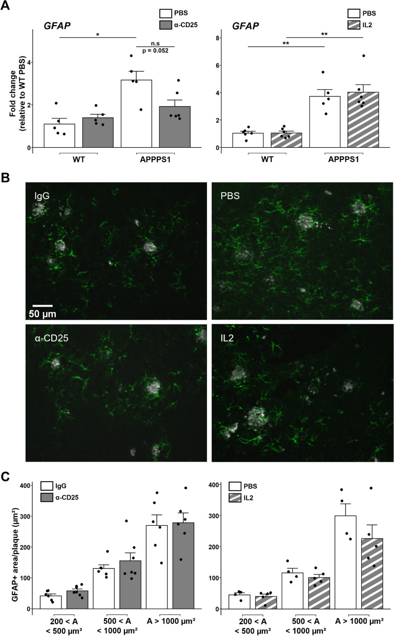
Fig. 1.
Modulation of Tregs does not alter plaque-associated global astrocyte reactivity. A Quantification of mRNA corresponding to the reactive astrocyte marker GFAP in the brain of mice treated with either PBS or anti-CD25 antibody (left), or with either PBS or IL-2 (right). Values were first normalized according to the expression of the housekeeping gene PPIA and then normalized to the mean value of PBS-injected WT control group. B Representative images of astrocytes (GFAP, green) associated to amyloid deposits (Aβ, white) in the cerebral cortex of mice treated with either isotypic control antibody (top left) or anti-CD25 (bottom left), or with either PBS (top right) or IL-2 (bottom right). C Quantification of GFAP immunoreactivity in close vicinity to amyloid deposits of different size ranges in the cerebral cortex of mice treated with either isotypic control antibody or anti-CD25 (left) or with either PBS or IL-2 (right). Results are represented according to the size of amyloid deposits that astrocytes are recruited towards. Mean +/− SEM (n > 20 amyloid deposits from 4–6 mice/ group). Mann–Whitney test: *P < 0.05; **P < 0.01; ***P < 0.001

