Fig. 2.
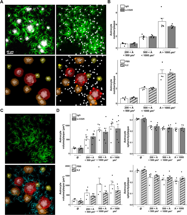
Modulation of Tregs does not affect the volume and sphericity of plaque-associated astrocytes. A Illustration of the 3D modeling process for analyzing astrocytes’ recruitment around amyloid deposits. Top left: representative raw image of astrocytes (GFAP, green) associated to cortical amyloid deposits (Aβ, white) from a 90-µm-thick cortical section. Top right: modeling of astrocytes’ soma (white). Bottom left: 3D-modeling of amyloid deposits (white) and their proximal environment (yellow to red, according to size range). Bottom right: representative image of astrocytes’ soma recruitment in the vicinity of amyloid deposits. Recruited astrocytes’ soma are represented as colored spheres according to the size of their corresponding amyloid deposit, unrecruited astrocytes’ soma are represented as white spheres. B Quantification of astrocyte recruitment in close vicinity to amyloid deposits of different size ranges in the cerebral cortex of APPPS1 mice treated for either the depletion (top) or amplification of Tregs (bottom). C Illustration of the 3D modeling process for analyzing astrocytes’ volume. Top: 3D modeling of astrocytes. Bottom: representative image of modeled astrocytes and amyloid plaques. Astrocytes in the vicinity of amyloid deposits are colored according to the size of their corresponding amyloid plaque, unrecruited astrocytes are represented in cyan. D Quantification of astrocytes’ volume (left) or sphericity (right) according to their proximity to amyloid deposits of different size ranges in the cerebral cortex of APPPS1 mice treated for either the depletion (top) or amplification of Tregs (bottom). Results are represented according to the size of amyloid deposits. Unrecruited astrocytes are referenced as Ø. Mean +/− SEM (n > 30 amyloid deposits from 4–6 mice/group). Mann–Whitney test: *P < 0.05; **P < 0.01; ***P < 0.001

