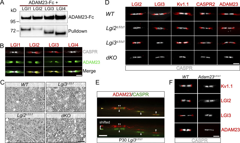
Figure 2.
LGI2 and LGI3 are ligands for ADAM23 at the JXP and play a role in localization of Kv1 channels in myelinated axons. (A) Western blot (WB) on tissue culture supernatants following co-transfection of HEK293T cells with ADAM23-Fc and one of the four LGI proteins. ADAM23-Fc (top) was precipitated from media with ProtA beads, co-precipitating interacting LGI proteins (bottom, pulldown). (B) IHC showing which of the LGI proteins are present at the JXP along with ADAM23. Adult WT teased sciatic nerve immunolabeled with CASPR (gray) LGI1, 2, 3, or 4 (red) and ADAM23 (green). Scale bar = 10 µm. (C) Semi-thin images of paraphenylenediamine-stained cross sections of sciatic nerves from WT, Lgi3Δ1/Δ1, Lgi2Δ1/Δ1, and double Lgi2Δ1/Δ1 and Lgi3Δ1/Δ1 knock-out (dKO) adult mice. Scale bar = 10 µm. (D) IHC on teased sciatic nerve fibers from P12 mice. WT tissue staining shown as a positive control (top) followed by images from mice lacking the expression of Lgi2 (Lgi2Δ1/Δ1), Lgi3 (Lgi3Δ1/Δ1), or both Lgi2 and Lgi3 (double knock-out - dKO). All images include the paranodal CASPR staining (gray) and one of the following: LGI2, LGI3, Kv1.1, CASPR2, and ADAM23 (red). Scale bar = 5 µm. (E) IHC on teased sciatic nerve from an adult (P30) Lgi3Δ1/Δ1 mouse, stained with antibodies against CASPR (green) and ADAM23 (red). Top image is a merge of both channels whereas bottom image represents split, horizontally shifted signals to indicate mislocalization of ADAM23 and its invasion of the paranodal region in the absence of LGI3 in an adult mouse. Scale bar = 10 µm. (F) Examination of LGI2 and LGI3 expression at the JXP in WT (left) and Adam23∆1/∆1 (right) P10 mice through IHC on teased sciatic nerve. All images show CASPR (gray) staining and Kv1.1, LGI2, LGI3, or ADAM23 (red). Scale bar = 5 µm. Source data are available for this figure: SourceData F2.

