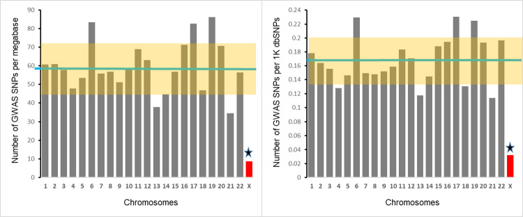
Fig 2. Chromosomal distributions of density (left panel) and fraction (right panel) of GWAS-detected SNPs.
Left panel depicts the number of GWAS-detected SNPs per megabase, right panel shows the number of GWAS-detected SNPs per thousand SNPs. Each bar represents a chromosome. Green horizontal line represents the mean for all autosomes. Highlighted area represents SD for autosomes. Star marks the significant difference between X-chromosome and the mean for autosomes.

