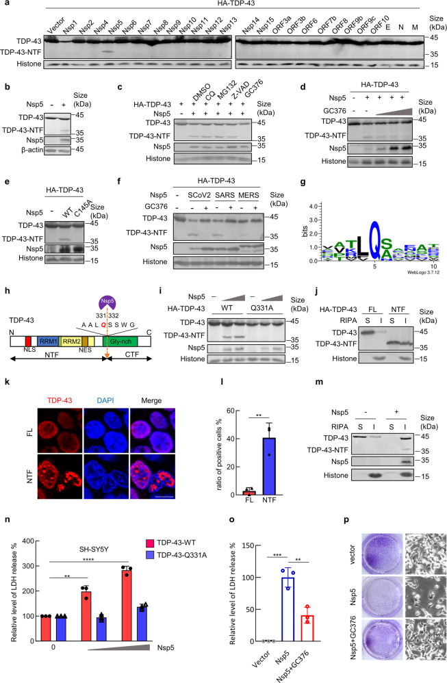
Fig. 1.
The main SARS-CoV-2 protease Nsp5 cleaves the TDP-43 protein into a cytotoxic form in human neural cells. a Expression plasmids of SARS-CoV-2-encoded proteins were cotransfected with pVR1012-HA-TDP-43 into HEK293T cells. Forty-eight hours later, samples were prepared for immunoblotting by using an anti-HA antibody. FL full length, NTF N-terminal fragment. b Immunoblotting data of endogenous TDP-43 cleaved by Nsp5. c Transfected HEK293T cells from (b) were treated with the indicated inhibitors. TDP-43 cleavage was detected by immunoblotting. CQ, chloroquine; Z-VAD, Z-VAD-FMK. DMSO was used in the solvent control group. d TDP-43 cleavage was inhibited by treatment with increasing concentrations of GC376 (0, 1, 5 or 20 μM). e HEK293T cells were cotransfected with pVR1012-HA-TDP-43 and pCAG-SARS-CoV-2-Nsp5-FLAG or its mutant Q145A. Samples were prepared for immunoblotting 48 h later. f Immunoblotting for TDP-43 cleaved by Nsp5 from SARS-CoV-2, SARS-CoV, and MERS-CoV. Transfected cells were treated with DMSO and GC376. g Logo analysis of the predicted cleavage site of SARS-CoV-2 Nsp5 by WebLogo3.7.12. h Schematic diagram of SARS-CoV-2-Nsp5 cleavage of TDP-43. NLS nuclear localization signal, NES nuclear export signal, RRM RNA recognition motif, Gly-rich glycine-rich domain. i The TDP-43 Q331A mutant is resistant to SARS-CoV-2 Nsp5. HEK293T cells were cotransfected with pVR1012-HA-TDP-43 or Q331A with increased expression vectors of SARS-CoV-2-Nsp5 (0, 100, and 200 nM). Cells were harvested at 48 h after transfection for immunoblotting assays. j Immunoblotting assays of the solubility of TDP-43 wild-type and NTF proteins. HEK293T cells were transfected as indicated and then collected after 48 h. Proteins were sequentially extracted using RIPA and 7 M urea buffers. S, RIPA soluble fraction; I, RIPA insoluble fraction. k Subcellular location of the indicated TDP-43 proteins. Scale bar, 10 μm. l The percentage of TDP-43 aggregates in cultures from (k). m Nsp5 decreases TDP-43 solubility. n SARS-CoV-2 Nsp5 enhances TDP-43 toxicity to SH-SY5Y human neuroblastoma cells. The release of LDH into the medium was used as an indicator of cytotoxicity. LDH levels were measured 72 h after cells were transfected with the indicated constructs. o GC376 relieved the cytotoxic effects of Nsp5 in human neuroblastoma cells. p Crystal violet staining (left panel) and bright-field (right panel) images of SH-SY5Y cells transfected with SARS-CoV-2 Nsp5 and treated with DMSO or 30 μM GC376. Scale bar, 50 μM. Error bars denote SEM; ANOVA test, n = 3 biologically independent experiments; ****p < 0.0001, ***p < 0.001, **p < 0.01

