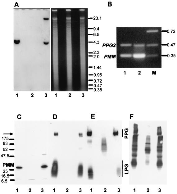
FIG. 4.
Analysis of L. mexicana wild type, a ΔPMM mutant, and a PMM gene addback mutant by Southern blotting, RT-PCR, and immunoblotting. (A) Southern blot analysis of PstI restriction enzyme-digested chromosomal DNA (10 μg) from L. mexicana wild type (lanes 1), a ΔPMM mutant (lanes 2). and a ΔPMM + cRIBPMM gene addback mutant (lanes 3). The digested DNAs were separated on an ethidium bromide-containing 0.7% agarose gel (right), blotted onto a nylon membrane, and incubated with a DIG-labeled PMM ORF probe (left). The sizes of DNA standards are indicated in kilobases. (B) Amplification of PMM mRNA from L. mexicana log-phase promastigote (lane 1) and amastigote (lane 2) by RT-PCR from total RNA. The loading was normalized to the coamplified cDNA fragment derived from the PPG2 gene, whose mRNA is approximately equally abundant in L. mexicana promastigotes and amastigotes (13). The sizes of DNA standards (lane M) are indicated in kilobases. (C to F) SDS-PAGE and immunoblotting of L. mexicana wild type and ΔPMM mutant total-cell lysates. Lanes 1, wild type; lanes 2, ΔPMM; lanes 3, ΔPMM + cRIBPMM. Each lane was loaded with 106 promastigotes (∼4 μg of protein). (C) Blot was probed with affinity-purified rabbit anti-L. mexicana PMM antibodies. The same or identically loaded blots were then stripped and probed with MAb LT6 (directed against [6Galβ1-4Manα1-PO4]x) (D), LT17(directed against [6(Glcβ1-3)Galβ1-4Manα1-PO4]x [x = unknown]) (E), and MAb L7.25 (directed against [Manα1-2]0-2Manα1-PO4) (F). The molecular masses and relative positions of standard proteins and the positions of PMM, LPG, and PPG are indicated. The arrow marks the border between stacking and separating gels.

