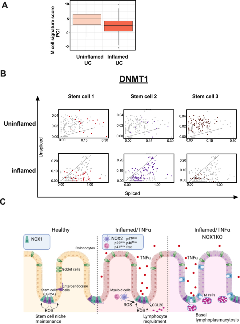
Figure 6. M cell signatures and DNMT1 RNA velocity correlated to UC inflammation status, implicating ROS-mediated mechanisms driving chronic inflammation.
A. Box whisker plot of UC patients’ M cell gene signature scores comparing bulk RNASeq (144 inflamed vs. 167 uninflamed rectal UC)-derived composite scores for PC1 (36.7% variance explained). Supplementary table 5 includes scores for the first 10 PCs. B. RNA velocity of DNMT1 in UC stem cells. C. Model for ROS-mediated, genotype-dependent differences in generating lymphoid aggregates, contributing to UC inflammation. i) Healthy: wild-type NOX1-mediated maintenance of the stem cell niche, ii) Inflamed/TNFα: expansion of CCL20+ cells generally, recruiting CCR6+ lymphocytes, and iii) Inflamed/TNFα/Nox1KO: ROS deficiency, together with TNFα treatment, induces M cells expansion and leads to basal lymphoplasmacytosis.

