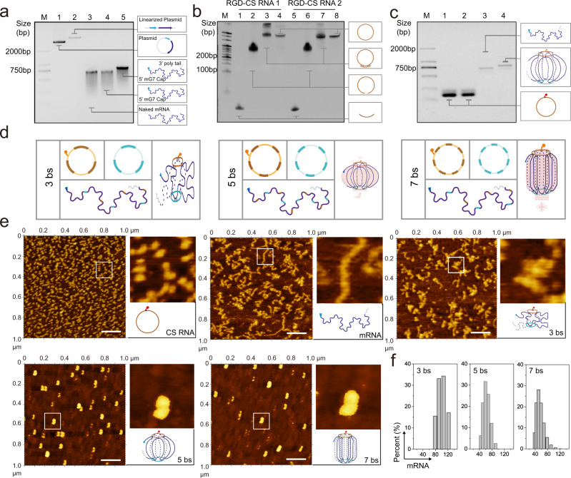
Fig. 2. Construction and characterization of Smad4 mRNA nano-lantern.
a–c Representative images from 3 independent experiments. Agarose gel electrophoresis of mRNA synthesis and modified (a), urea-polyacrylamide gel electrophoresis of circular RNA staples (5 bs) synthesize (b), and formation of Smad4 mRNA nano-lantern (c). (d) Possible structures of the Smad4 mRNA nano-lantern with different binding sites. (e) Representative AFM imaging of naked Smad4 mRNA and different forms of mRNA nano-lantern (with 3, 5, and 7 binding sites) from 3 independent experiments, Scale bar: 0.2 μm. (f) Diameter of different forms of mRNA nano-lantern. Panel (c–e) created with BioRender.com. Source data are provided as a Source Data file.

