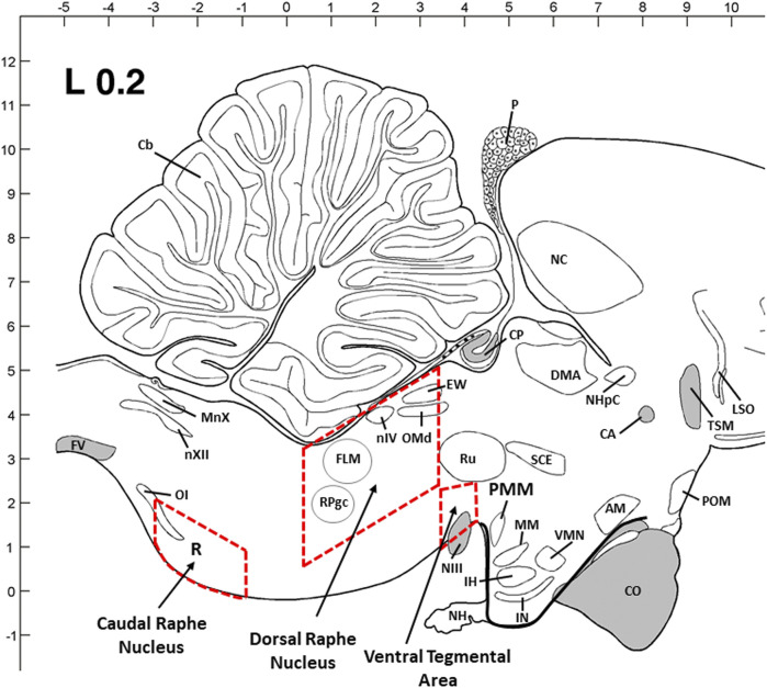
FIGURE 5.
Sagittal view of three dissection areas of the chicken brain [dorsal raphe nucleus (DRN) and caudal raphe nucleus (CRN) of the brainstem and ventral tegmental rea (VTA) of the midbrain]. Dimensions of the dissected tissues are coronal with 2.5–3 mm (W) x 1–1.5 mm (H) x 2.5–3.0 mm (L) for DRN, 2–2.5 mm (W) x 1–1.2 mm (H) x 2.5–3.0 mm (L) for CRN, and 3–3.5 mm (W) x 2–3 mm (H) x 1–1.2 mm (L) for VTA. The thickness (W, H, and L) was adjusted proportionally from young birds to older birds based on the brain size and structure. Abbreviations: AM: anterior medial hypothalamic nucleus; CA: anterior commissure; Cb: cerebellum; CO: optic chiasma; CP: posterior commissure; DMA: dorsomedial nucleus; EW: Edinger–Westphal nucleus; FLM: medial longitudinal fasciculus; FV: ventral fasciculus; IH: inferior hypothalamic nucleus; IN: infundibular hypothalamic nucleus; LSO: lateral septal organ; MM: medial mammillary nucleus; MnX: nucleus motorius dorsalis nervi vagi; NC: caudal neostriatum; NH: neurohypophysis; NHpC: nucleus of the hippocampal commissure; NIII: oculomotor nerve; nIV: trochlear nerve nucleus; nXII: hypoglossal nerve nucleus; OI: inferior olivary nucleus; OMd: dorsal oculomotor nucleus; P: pineal gland; POM: medial preoptic nucleus; PVN: paraventricular nucleus; RPgc: nucleus of caudal pontine reticular gigantocellular; Ru: red nucleus; SCE: stratum cellular externum; TSM: septopallio-mesencephalic tract; VMN: ventromedial hypothalamic nucleus.

