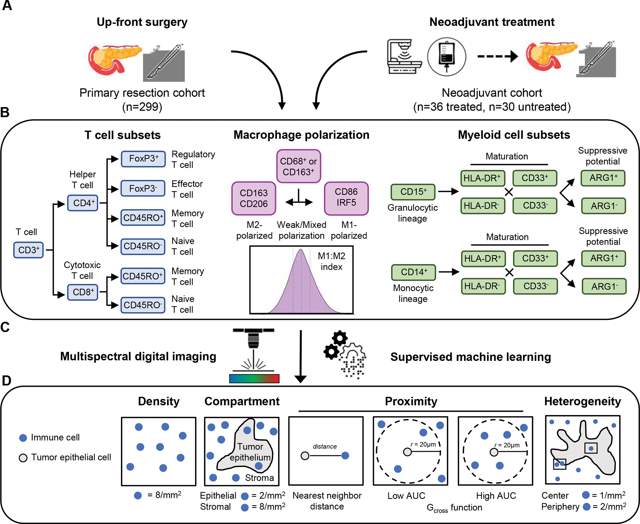
Figure 1. Overview of study cohort and analysis approach.

Two pancreatic ductal adenocarcinoma tissue cohorts were analyzed, including a primary resection cohort and a neoadjuvant-treatment cohort (A). T cell subsets (helper and cytotoxic T cells, including regulatory activity and naïve/memory status), differentially-polarized macrophages (M1- and M2-polarized macrophages, including strength of polarization) and myeloid cell subsets (granulocytes and monocytes, including maturity and ARG1 immunosuppressive activity) were evaluated using three multiplexed immunofluorescence panels (B). Combinatorial protein expression and cytomorphology data were analyzed by multispectral digital image analysis and supervised machine learning to identify specific cell types (C). Quantification of immune cell abundance was performed to measure immune cell densities overall and within separate tissue compartments (tumor epithelium or stroma); immune cell proximity to tumor cells was assessed by nearest neighbor distance (NND) and the Gcross function; and regions of interest were analyzed to assess immune cell heterogeneity (D).
