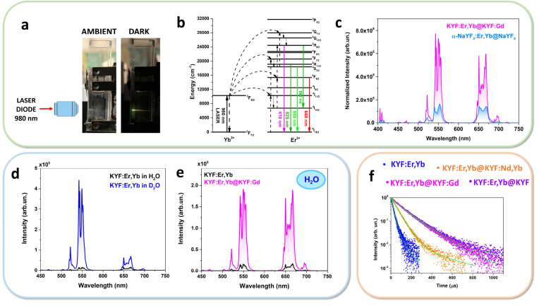
Figure 3.
(a) Snapshots of the UC emission for a colloidal dispersion of KYF:Er,Yb@KYF:Gd (concentration of 20 mg/mL). (b) Energy level scheme for Yb3+ and Er3+ ions and UC mechanisms. (c) UC emission spectra of KYF:Er,Yb@KYF:Gd and α-NaYF4:Er,Yb@NaYF4 in colloidal dispersion, normalized on Yb3+ concentration, under 980 nm laser excitation, P ∼ 4 kW cm–2. (d) UC spectra of colloidal dispersions of core KYF:Er,Yb (20 mg/mL) in H2O and D2O. (e) UC spectra (λexc = 980 nm) for colloidal dispersions of core KYF:Er,Yb (20 mg/mL) and core@shell KYF:Er,Yb@KYF:Gd (20 mg/mL) in H2O. (f) Emission decays in the green due to the 2H11/2, 4S3/2 →4I15/2 transitions of Er3+ (λexc = 980 nm, λem = 543 nm) for all the nanostructures (concentration of 20 mg/mL).

