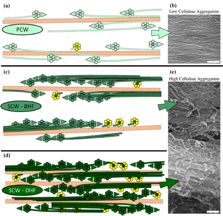
Figure 4.
Models and scanning electron microscope images depicting the aggregation of cellulose microfibrils in PCW (a) and (b) and in SCW before early formation (BHF) (c) and (e) and during late formation (DHF) (d) and (e); scale bars = 200 nm. Reproduced with permission from ref (44) (CC-BY).

