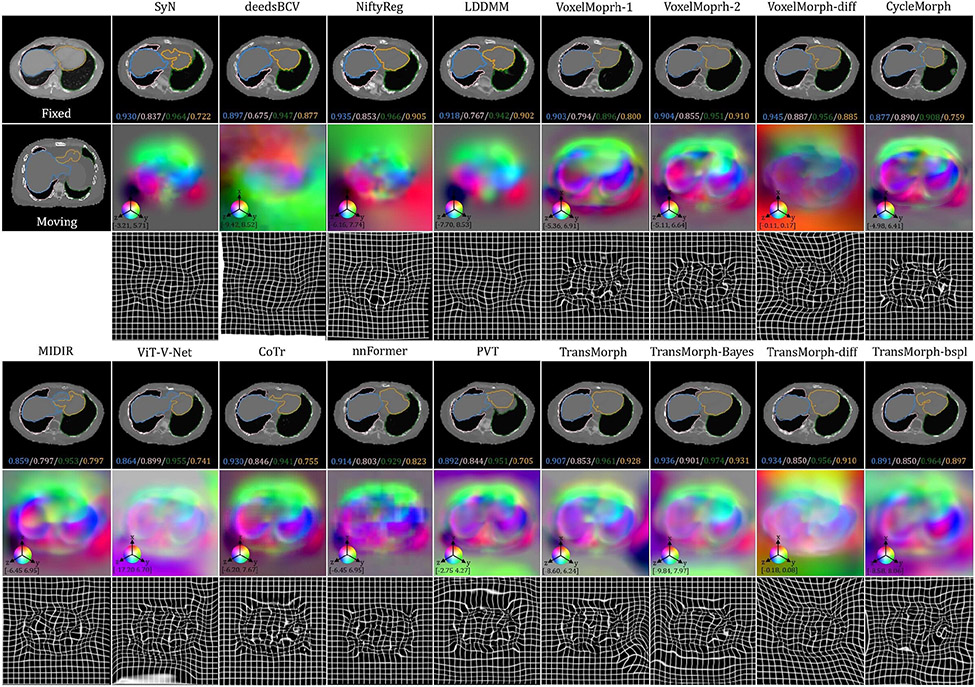
Fig. E.26:
Additional qualitative comparison of various registration methods on the XCAT-to-CT registration task. The first row shows the deformed moving images, the second row shows the deformation fields, and the last row shows the deformed grids. The spatial dimension x, y, and z in the displacement field is mapped to each of the RGB color channels, respectively. The [p, q] in color bars denotes the magnitude range of the fields.

