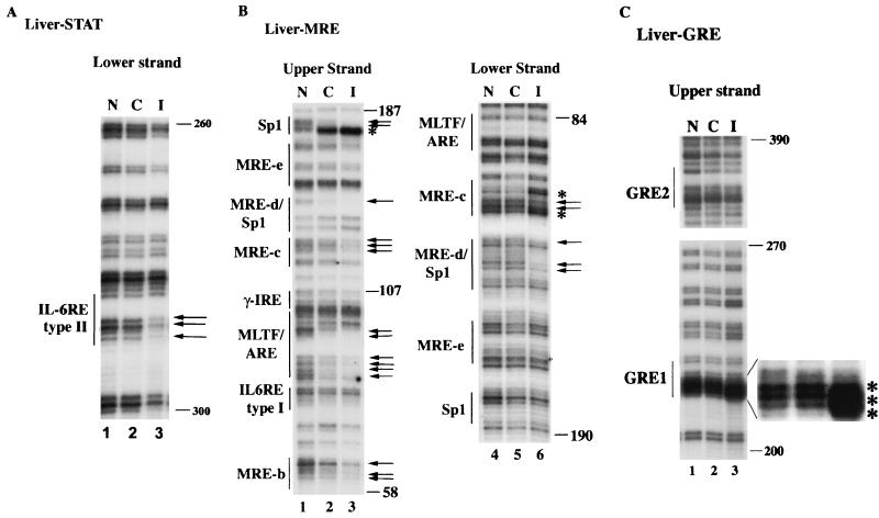
FIG. 4.
IVGF studies reveal protection of IL-6 type II RE, MREs, and GRE1 of the MT-I promoter in the livers of infected C57BL/6 mice. The nuclei isolated from the livers of control and infected mice suspended in PBS, as well as naked DNA, were treated with DMS. DNA was then purified, and methylated guanines were cleaved with piperidine. An identical amount of DNA (2 μg) from each sample was then subjected to LM-PCR with specific sets of primers (described in Materials and Methods). The G ladder generated was separated on a sequencing gel and analyzed by autoradiography. (A) Footprinting of the upper and lower strands of MT-I promoter spanning IL-6 RE with 5′ STAT3 primers. (B) Footprinting of the promoter region spanning MREs with 3′ and 5′ mMT-I primers. (C) Footprinting of the promoter region upstream of MT-II spanning GREs with 3′ GRE primers. Lanes N, C, and I denote LM-PCR with naked DNA and DNA isolated from the liver nuclei of uninfected and infected animals, respectively. Numbers on the right denote positions of G residues upstream of the +1 site.

