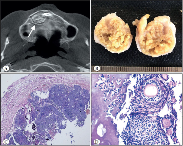
Figure 1.
Adenomatoid odontogenic tumor. A) Axial CBCT view of right maxillary unerupted canine region showing well-defined lesion with visible internal mineralization (arrow). B) Macroscopic appearance of the same case; rounded masses showing a solid yellowish pattern on the cut surface. C) Tumor demonstrating a fibrous capsule with odontogenic epithelium in solid nodules (H&E; x40). D) At high power, duct-like structures and calcifications clearly seen (H&E; x200).

