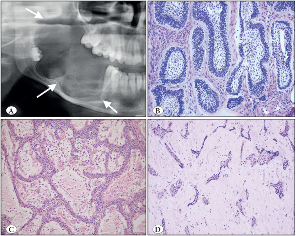
Figure 3.
Ameloblastoma. A) Cropped panoramic radiograph showing a typical expansive, multilocular radiolucency (arrows). B) Follicular pattern; islands where peripheral cells show hyperchromatic nuclei in a palisading pattern, reserve polarity and looser stellate reticulum-like or squamous change in the center (H&E; x200). C) Plexiform pattern; anastomosing cords and strands of epithelium (H&E; x100). D) Desmoplastic pattern; epithelial islands in dense stroma (H&E; x100).

