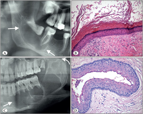
Figure 10.
Orthokeratinized odontogenic cyst (A-B) vs. Odontogenic keratocyst (C-D). A) Cropped panoramic radiograph showing a well-circumscribed unilocular radiolucency associated with an unerupted third molar (arrows). B) OOC is lined by a uniform stratified squamous epithelium with orthokeratosis, prominent granular cell layer and bland, unpalisaded basal cells (H&E; x200). C) Cropped panoramic radiograph showing a multilocular radiolucency of the left mandibular body and ramus (arrows). D) OKC is lined by a uniform stratified squamous epithelium with a corrugated surface of parakeratin and palisaded and hyperchromatic basal cells (H&E; x200).

