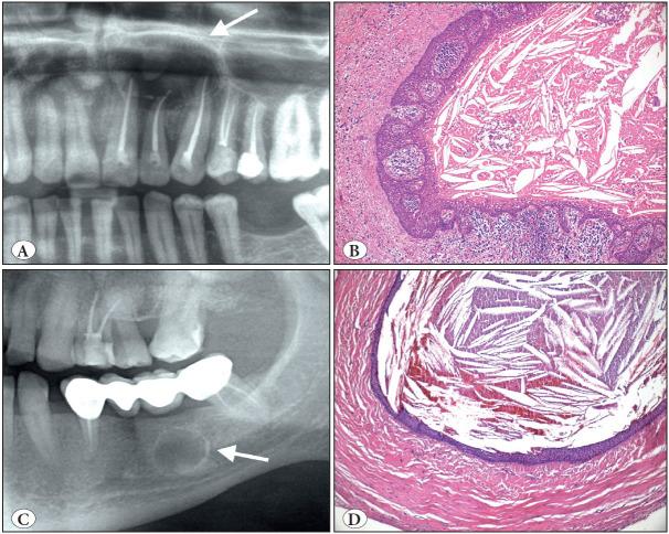
Figure 9.
Radicular cyst. A) Cropped panoramic radiograph showing a well-defined, corticated unilocular radiolucency at the apices of endodontically treated teeth (arrow). B) Lining by non-keratinized stratified squamous epithelium with epithelial hyperplasia in a characteristic arcading pattern. Cyst wall is inflamed (H&E; x100). C) Cropped panoramic radiograph of residual cyst showing a well-circumscribed, corticated unilocular radiolucency in an edentulous area of the left mandible (arrow). D) Residual (or long-standing) cyst showing less inflamed wall and a more regular thin epithelium (H&E; x100).

