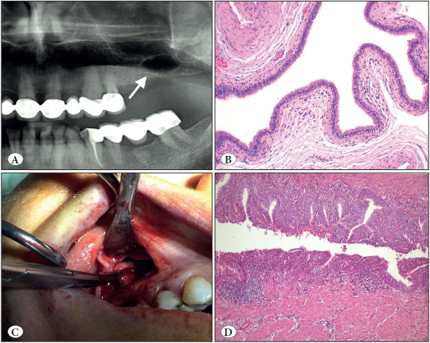
Figure 13.
Surgical ciliated cyst. A) Cropped panoramic radiograph showing a well-demarcated unilocular radiolucency of the left maxilla with a history of traumatic tooth extraction (arrow). B) The cyst lined entirely by respiratory epithelium (H&E; x100). C) Intra-operative view of the case located right site of maxilla. D) This case shows hyperplastic pseudostratified ciliated columnar epithelium with mucous cells and inflamed cyst wall (H&E; x200). Awareness of this entity prevents misdiagnosis.

