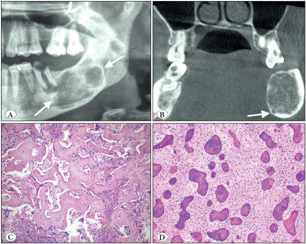
Figure 5.
Cemento-ossifying fibroma. A) Cropped panoramic radiograph showing a well-defined, expansile radiolucency in the posterior mandible (arrows). B) Coronal CBCT view showing the expansion and displacement of the inferior mandibular canal (arrow). C) COF is a prototype benign fibro-osseous jaw lesion. The matrix produced can be trabecular with cellular inclusions and osteoblastic rimming like bone (H&E; x200) or D) COF often contains smaller rounder and acellular matrix similar to cementum (H&E; x200).

