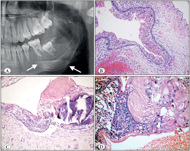
Figure 11.
Calcifying odontogenic cyst. A) Cropped panoramic radiograph showing well-defined, unilocular, mixed radiolucent/radiopaque lesion with distinct cortical expansion of the left posterior mandible and ramus (arrows). B) Low power shows a cystic architecture with prominent eosinophilic, polyhedral cells (ghost cells). (H&E; x200). C) Focus of ghost cells, some of which show calcification (H&E; x200). D) Characteristic ghost cells where the nucleus is lost but cytoplasmic outlines are maintained (H&E; 400).

