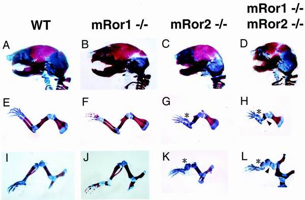
FIG. 3.
Examination of craniofacial bones and appendicular skeletons. The craniofacial bones and appendicular skeletons from wild-type (WT), mRor1−/−, and mRor2−/− newborns and double mutant embryos (E19.5) were double stained with Alizarin red and Alcian blue as described in Materials and Methods. Lateral views of the skeleton of the head (A to D) and extremities (forelimb [E to H] and hind limb [I to L]) from wild-type and mutant newborns and a double mutant embryo are shown. Asterisks and arrowheads, dysplasia of the distal and proximal parts of limb bones in mRor2−/− and double mutant mice, respectively. In some cases, apparently delayed ossification of the cranial suture was also observed (data not shown).

