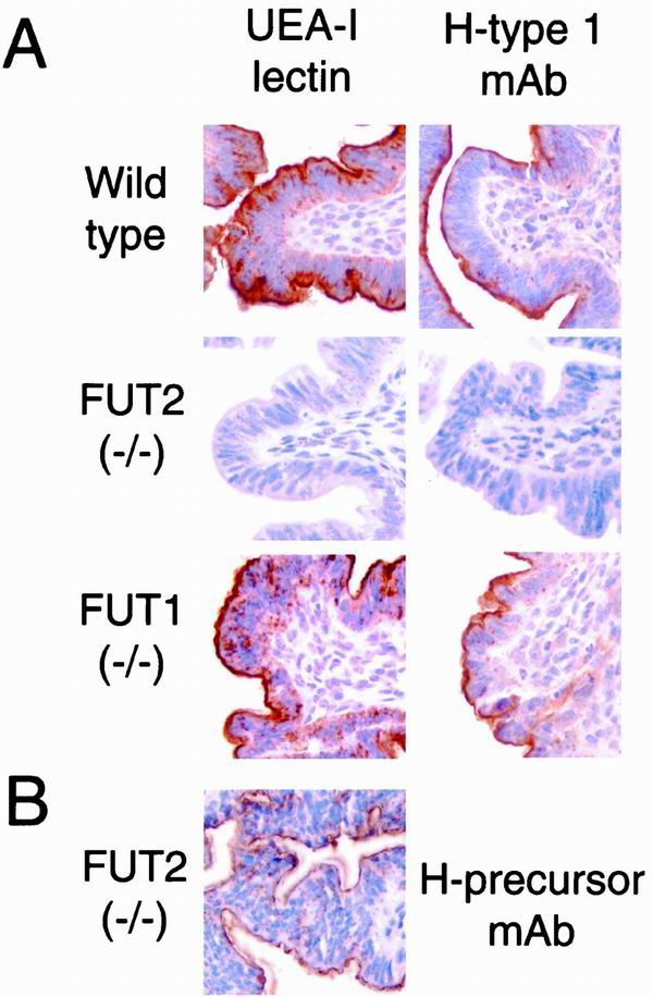
FIG. 3.
Lectin and blood group immunohistochemistry of the uterus in wild-type, FUT2 null, and FUT1 null mice. Adult female mice were sacrificed on the first day of estrus and processed for immunohistochemistry (see Materials and Methods). (A) Uteri stained with the α(1,2)fucosylated glycan-specific lectin UEA-I or H type I-specific MAb. The brown staining identifies H antigen-reactive glycans within the glycocalyx on the apical surfaces of luminal uterine epithelial cells from wild-type mice and FUT1−/− mice. Staining is absent from the glycocalyx of FUT2−/− uterine epithelia. (B) A FUT2−/− uterus was stained with a MAb (H-precursor MAb; BG-1) specific for nonfucosylated type I glycans that serve as precursors for H type I blood group synthesis (see Materials and Methods).

