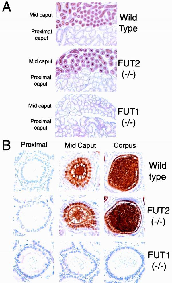
FIG. 4.
Lectin histochemistry of the epididymis in wild-type, FUT2 null, and FUT1 null mice. Epididymides from adult wild-type, FUT2 null, and FUT1 null male mice were stained with UEA-I (see Materials and Methods). (A) Low magnification (×50) of epididymis UEA-I histochemistry. UEA-I staining is prominent in the mid-caput but absence from the proximal caput in wild-type mice and FUT2−/− mice. UEA-I does not stain the mid-caput or proximal caput in FUT1−/− mice. (B) High magnification (×500) of epididymis UEA-I histochemistry. Single epididymal tubules are shown from proximal caput, mid-caput, and corpus from adult wild-type, FUT2 null, and FUT1 null mice. Mid-caput epithelial cells and adjacent spermatozoa show specific UEA-I staining in wild-type and FUT2 null mice but not FUT1 null mice. Spermatozoa from the corpus epididymis display dense UEA-I staining in wild-type and FUT2 null mice but not FUT1 null mice.

