Wang et al. 10.1073/pnas.0502917102. |
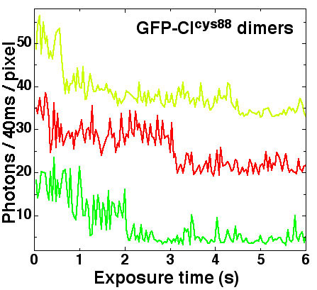
Fig. 12.
Staggered fluorescence time traces of single covalent GFP-cICys-88 dimers taken from 3.7-Hz pulsed-illumination measurements with an exposure time of 40 ms. Peak photon counts from the protein dots were used. The illumination intensity was 150 W/cm2.
Fig. 13
. The number of GFP-LacI bound to lacO256-DNA with and without oxygen scavenging. The two histograms are identical, indicating the absence of quenching between GFP and BOBO-3.