Kim et al. 10.1073/pnas.0505622102. |

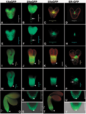
Fig. 7. Movement pattern of soluble GFP during embryo development.

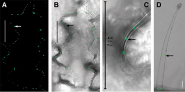
Fig. 8. Subcellular localization of Tobacco mosaic virus (TMV) P30-1´ GFP in transgenic tobacco leaves.

Fig. 9. Movement pattern of Tobacco mosaic virus (TMV) P30-GFP during embryo development.