Qiao et al. 10.1073/pnas.0502137102. |
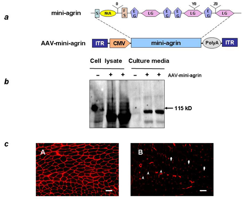
Fig. 7. Construction and characterization of mouse miniagrin gene and expression vector. (a) Construction of mouse miniagrin gene and AAV vector. The miniagrin contains the signal sequence (s), the N-terminal agrin (NtA) domain that binds to laminin, one follistatin-like domain (FS), and the C terminus containing the EGF-like domain (EG) and laminin G-like domains (LG) that bind to a-dystroglycan. The miniagrin gene (GenBank accession no. AY914875) was cloned in AAV vector between a CMV promoter and a bovine growth hormone poly(A) sequence. The entire vector DNA is flanked by the AAV inverted terminal repeats (ITR). (b) Western analysis of AAV-miniagrin gene expression and protein secretion in cell culture. (c) IF staining of AAV2-miniagrin gene expression after intramuscular injection in tibialis anterior muscle of normal mouse. (A) Overexpression and correct extracellular localization of the miniagrin is observed 4 weeks after vector injection. (B) The uninjected normal control muscle shows punctuated agrin staining at the neuromuscular junctions (arrowheads) and blood vessels (arrows). (Scale bars: 100 mm.)
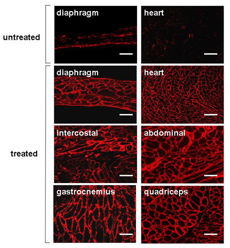
Fig. 8. IF staining of agrin on muscle cross sections showed overexpression of miniagrin in multiple muscle groups and heart. The AAV1 vector was injected i.p. in dyw/dyw neonates, and the mice were killed at 4 months of age. (Scale bars: 100 mm.)
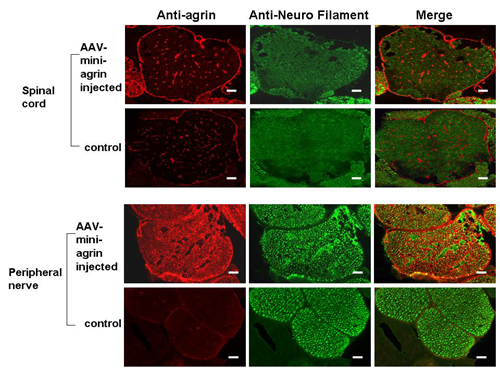
Fig. 9. Miniagrin protein expressed in the peripheral nervous system but not in the spinal cord by AAV injection. Agrin is stained as red, and the neuron filament is stained as green. There was no difference of agrin expression between the treated and the untreated spinal cord, although agrin was significantly expressed in the peripheral nerve roots. (Scale bars: 100 mm.)