Ewers et al. 10.1073/pnas.0504407102. |
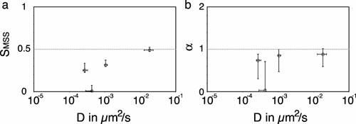
Fig. 6. Comparison of the relative position and error of representative trajectories in plots of D vs. SMSS (a) and D vs. a (b), with a = g2. The errors in the x and y direction are the 1-s intervals as calculated in ref. 1 (details are in Supporting Materials and Methods).
1. Qian, H., Sheetz, M. P. & Elson, E. L. (1991) Biophys. J. 60, 910–921.
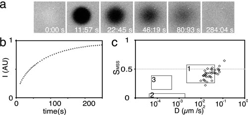
Fig. 7. VLPs bound to artificial lipid bilayers containing GD1a are highly mobile. (a) Laser scanning confocal micrograph time series of a fluorescence recovery after photobleaching experiment performed on lipid bilayers of dioleoylphosphatidylcholine containing fluorescein-dipalmitoylphosphatidylethanolamine and unlabeled GD1a. (b) Micrographs of a representative experiment (bleach region diameter, 45 mm) and the plot of the fluorescence recovery over time are shown. (c) D/SMSS plot of virus-like particle trajectories recorded by total internal reflection fluorescence microscopy while bound to lipid bilayers containing GD1a.
Movie 1. Alexa Fluor 568 (AF568)-labeled murine polyoma virus-like particles (VLPs) bound to a 3T6 cell. Epi-fluorescence of AF568-labeled VLPs was recorded at 0.5 Hz and the movie is played at 10 times real time. The field recorded is 23 ´ 19 mm in size.
Movie 2. Binding of a virus-like particle (VLP) to a 3T6 cell recorded by total internal reflection fluorescence microscopy. The binding event was recorded 15 min after addition of VLPs to the cells on the stage of the microscope. The field is 2 ´ 1.68 mm, and the movie was recorded at 20 Hz and is played in real time.
Movie 3. Fluorescence recovery after photobleaching time series of confocal micrographs of fluorescein-dipalmitoylphosphatidylethanolamine in an artificial lipid bilayer. A circular region with a 45-mm diameter was photobleached with the 488-nm line of a Zeiss LSM 510 confocal laser scanning microscope, and the fluorescence signal was recorded at increasing time intervals to measure recovery of fluorescence intensity. The time scale is nonlinear because of the animation of the time series.
Movie 4. Single-particle tracking of virus-like particles bound to an artificial lipid bilayer containing GD1a recorded by total internal reflection fluorescence microscopy. Single-particle tracking was performed after fluorescence recovery after photobleaching experiments on the same bilayer that confirmed continuity and fluidity of the bilayer. The movie is recorded at 20 Hz and is sized at 12 ´ 12 mm and played in real time.
Movie 5. Moving window analysis of the trajectory of a virus-like particle followed from the moment of binding in a D/SMSS plot. For illustration, the trajectory is plotted next to the moving window plot. The point of the trajectory plotted always corresponds to the first of the 120 frames analyzed in the respective moving window.
Movie 6. Total internal reflection fluorescence movie of clathrin light-chain–GFP-expressing 3T6 cell with several bound Alexa Fluor 568-labeled virus-like particles (VLPs). The movie is recorded at 10 Hz 45 min after addition of VLPs to live cells on stage. The recorded field is 12 ´ 12 mm, and the movie is played in real time.
Movie 7. Total internal reflection fluorescence movie of caveolin-1 GFP-expressing 3T6 cell with several bound Alexa Fluor 568-labeled virus-like particle (VLPs). The movie is recorded 45 min after addition of VLPs to live cells on stage at 10 Hz. The recorded field is 12 ´ 12 mm, and the movie is played in real time.
Movie 8. Total internal reflection fluorescence movie of b-actin GFP-expressing 3T6 cell after addition of 0.2 mM latrunculin A. The movie starts with the addition of the drug and is recorded at 0.1 Hz. The recorded field is 42.4 ´ 22.4 mm.