Liu et al. 10.1073/pnas.0511342103. |
Supporting Figure 6
Supporting Figure 7
Supporting Figure 8
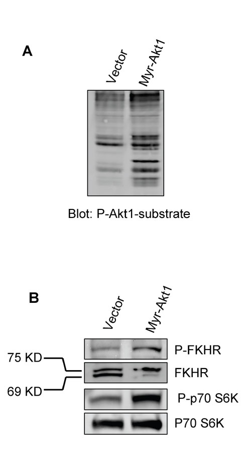
Fig. 6. Effect of myr-Akt1 expression on protein phosphorylation. (A) Increased phosphorylation of Akt1 substrates in myr-Akt1 transfectants. (B) Specific phosphorylation of FKHR and p70S6K by myr-Akt1.

Fig. 7. Myr-Akt1 expression enhances cell proliferation and decreases apoptosis in cells grown in 3D lrECM. (A) myr-Akt1 significantly increased the percentage of Ki-67-positive cells at days 2 and 4. (B) myr-Akt1-expressing cells showed significantly decreased TUNEL staining at days 6, 8, and 10.

Fig. 8. Expression of TSC2 increases stress fibers and focal adhesion in cells expressing myr-Akt1. Actin stress fibers (Left) and paxillin staining of focal adhesions (Right) are decreased in myr-Akt1-expressing cells (Middle) as compared with vector-transformed cells (Top). Coexpression of TSC2 with myr-Akt1 (Bottom) reestablishes stress fibers and focal adhesions. (Scale bar, 20 m m.)