Supplementary material for Ohnishi et al. (2001) Proc. Natl. Acad. Sci. USA 98 (2), 670–675. (10.1073/pnas.021546498)

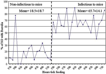
Fig. 9.
Borrelia burgdorferi infection of tick salivary glands. Infected ticks were allowed to feed on mice and the ticks were removed from individual mice at 2-hr intervals starting at 37 hr and ending at 72 hr into the blood meal. The tick salivary glands were dissected out and stained with a FITC-conjugated polyclonal Borrelia antibody as described in Materials and Methods. The data were analyzed to determine the proportion of infected salivary glands at each 2-hr interval. The vertical dashed line separates ticks that had attached to the host for too short a time to infect mice (<53 hr) from those that had attached long enough to infect mice (>53 hr) (see Fig. 2 for mouse transmission data).