Supplementary material for Ohnishi et al. (2001) Proc. Natl. Acad. Sci. USA 98 (2), 670–675. (10.1073/pnas.021546498)

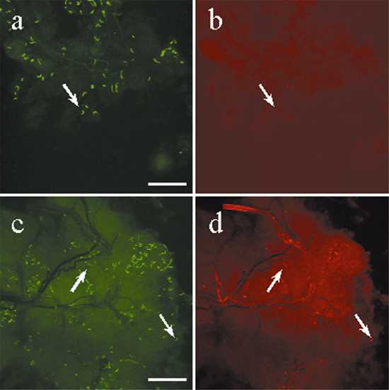
Fig. 10.
Direct dual immunofluorescence of Borrelia burgdorferi in tick salivary glands. Ticks were allowed to feed on mice for 67-69 hr, after which the salivary glands were removed and used in DFA experiments. (a and b) The same field stained with a FITC-conjugated polyclonal B. burgdorferi antibody (a) and a Texas Red-conjugated mAb against OspA (b). (c and d) The same field stained with a FITC-conjugated polyclonal B. burgdorferi antibody (c) and a Texas Red-conjugated mAb against OspC (d). (Bars = 100 mm.)